
|

|
Pictures Gallery

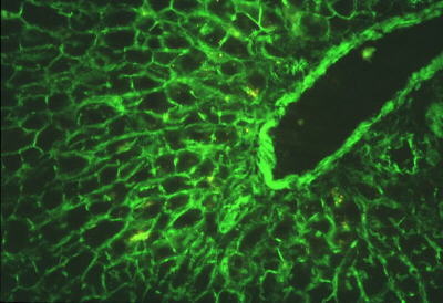
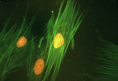
Mouse liver: Hoechst + Alexa 488 Phalloidin HEP-2 cells after a 30 min. pulse of BrdU: FITC labelled Anti-BrdU + Trypan Blue counterstaining Rat liver- Alexa 488 Phalloidin Human smooth muscle cells: FITC labelled Anti-smooth muscle cell alpha actin + propidium iodide counterstaing for DNA
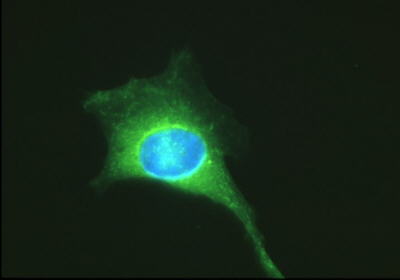
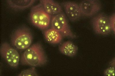
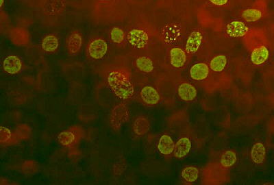
HEP-2 cells: FITC labelled Anti-Ki-67 + Trypan Blue counterstaining Human fibroblasts: FITC labelled Anti-ribonucleotide reductase (M1 subunit + Hoechst 33258 counterstaining for DNA)
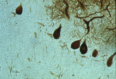
Rat cerebellum (17 days) after immunoreaction for Calbindin.
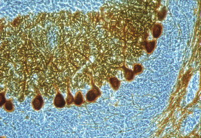
Phase contrastMonkey adult cerebellum (Macaca fascicularis) treated with MPTP after immunoreaction fot Calbindin.
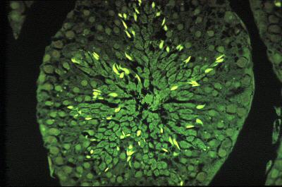
Differential Interference Contrast (DIC)Mouse seminiferous tubules, cross section: thiol groups of sperm protamins were stained with fluorescein mercuric acetate Apoptotic HEP-2 cells stained with TUNEL technique: FITC labelled DNA precursors were used, and sample was counterstained with Trypan Blue
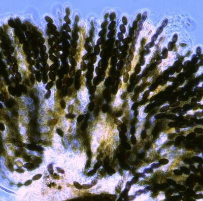
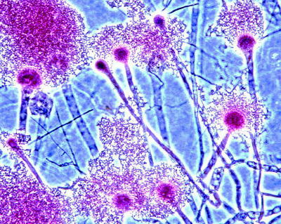
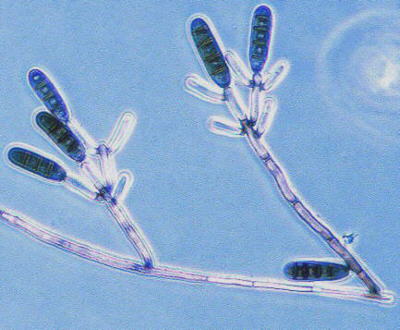
1) Acanthophiobolus sp. 2) Sordaria sp.: asci and ascospores 3) Aspergillus fumigatus: conidia and conidiophores with uniseriata sterigmata 4) Dendryphiopsis atra: conidiophores and conidia


Paramecium primaurelia - DIC
Below, the same field observed in fluorescence microscopyFluorescence microscopy + DIC
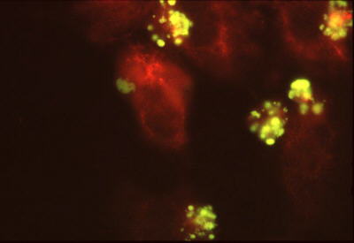
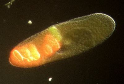
Paramecium primaurelia first fed with BSA-FITC and then with BSA-Texas red. Red-dyed food vacuoles utilizing the retrieved membrane from green-dyed food vacuoles are yellow-labeledParamecium primaurelia first fed with BSA-FITC and then with BSA-Texas red:
phagocytotic vacuoles are green or red labeled respectivelyFluorescence microscopy + DIC
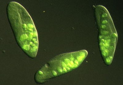
Paramecium primaurelia fed with BSA-FITC: phagocytotic vacuoles are green labeled
With the collaboration of:

A CN Biosciences Company by


This site is created and maintained by Dr. Vittorio Bertone