|
|
|
|
BASICS IN FLUORESCENCE MICROSCOPY AND FLUOROCHROMES (November 26-27, 1998) |

|

|
|
University of Pavia
Department of Animal Biology
Laboratory of Comparative Anatomy
and
Laboratory of Cell Biology
Piazza Botta 10, 27100 Pavia, Italy
| Prof. Isabel Freitas
Laboratorio di Anatomia Comparata Dipartimento di Biologia Animale Università di Pavia Piazza Botta 10, 27100 Pavia, Italy phone: + 39 - 0382 - 506317 fax: + 39 - 0382 - 506406 e-mail: freitas@unipv.it |
Prof. Carlo Pellicciari
Laboratorio di Istologia ed Antropologia Departimento di Biologia Animale Università di Pavia Piazza Botta 10, 27100 Pavia, Italy phone: + 39 - 0382 - 506420 fax: + 39 - 0382 - 506325 e-mail: pelli@unipv.it |
| Dr. Heinz Gundlach
Carl Zeiss Carl Zeiss Str. 1 D-7347 Oberkochen, Germany phone: + 49 - (0)7364 - 202071 fax: + 49 - (0)7364 - 204013 e-mail: gundlach@zeiss.de |
|
| Dr. Thomas Beller
Carl Zeiss S.p.A. Viale delle Industrie, 18 20020 Arese (Mi), Italy phone: + 39 - 02 - 937731 fax: + 39 - 02 - 93773301 e-mail: micro@zeiss.it |
Dr. Alessandro Borsatti
Carl Zeiss S.p.A. Viale delle Industrie, 18 20020 Arese (Mi), Italy phone: + 39 - 02 - 937731 fax. + 39 - 02 - 93773301 e-mail: micro@zeiss.it |
| Dr. Giovanni Bottiroli
Centro di Studio per lIstochimica del C.N.R. Piazza Botta 10, 27100 Pavia, Italy phone: + 39 - 0382 - 506412 fax: + 39 - 0382 - 506430 e-mail: botti@dragon.ian.pv.cnr.it |
Dr. Heinz Gundlach
Carl Zeiss Carl Zeiss Str. 1 D-7347 Oberkochen, Germany phone. + 49 - (0)7364 - 202071 fax: + 49 - (0)7364 - 204013 e-mail: gundlach@zeiss.de |
| Dr. Giuliano Mazzini
Centro di Studio per lIstochimica del C.N.R. Piazza Botta 10, 27100 Pavia, Italy phone: + 39 - 0382 - 506266 fax: + 39 - 0382 - 506430 e-mail:mazzi@dragon.ian.pv.cnr.it |
Prof. Sergio Barni
Laboratorio di Anatomia Comparata Dipartimento di Biologia Animale Università di Pavia Piazza Botta 10, 27100 Pavia, Italy phone: + 39 - 0382 - 506421 fax: + 39 -0382 - 506406 e-mail: anatcomp@unipv.it |
| Dr. Luigi Sciola
Dipartimento di Scienze Fisiologiche, Biochimiche e Cellulari Università di Sassari Via Muroni 25, 07100 Sassari, Italy phone: + 39 - 079 - 228651 fax: + 39 - 079 - 228615 e-mail: sciola@ssmain.uniss.it |
| Dr. Luigi Sciola
Dipartimento di Scienze Fisiologiche, Biochimiche e Cellulari Università di Sassari Via Muroni 25, 07100 Sassari, Italy phone: + 39 - 079 - 228651 fax: + 39 - 079 - 228615 e-mail: sciola@ssmain.uniss.it |
Dr. Vittorio Bertone
Laboratorio di Anatomia Comparata Dipartimento di Biologia Animale Università di Pavia Piazza Botta 10, 27100 Pavia, Italy phone: + 39 - 0382 - 506317 fax: + 39 - 0382 - 506406 e-mail: bertone@unipv.it |
| Dr. Patrizia Griffini
Laboratorio di Anatomia Comparata Dipartimento di Biologia Animale Università di Pavia Piazza Botta 10, 27100 Pavia, Italy phone: + 39 - 0382 - 506317 fax: + 39 - 0382 - 506406 e-mail: freitas@unipv.it |
Dr. Barbara Bono
Istituto di Patologia Generale C. Golgi Università di Pavia Piazza Botta 10, 27100 Pavia, Italy phone: + 39 - 0382 - 506334 fax: + 39 - 0382 - 303673 e-mail: golgi@unipv.it |
| Dr. Simona Fracchiolla
Laboratorio di Anatomia Comparata Dipartimento di Biologia Animale Università di Pavia Piazza Botta 10, 27100 Pavia, Italy phone: + 39 - 0382 - 506317 fax: + 39 -0382 - 506406 e-mail: freitas@unipv.it |
Dr. Alessandra Spano
Dipartimento di Scienze Fisiologiche, Biochimiche e Cellulari Università di Sassari Via Muroni 25, 07100 Sassari, Italy phone: + 39 - 079 - 228651 fax: + 39 - 079 - 228615 e-mail: sciola@ssmain.uniss.it |
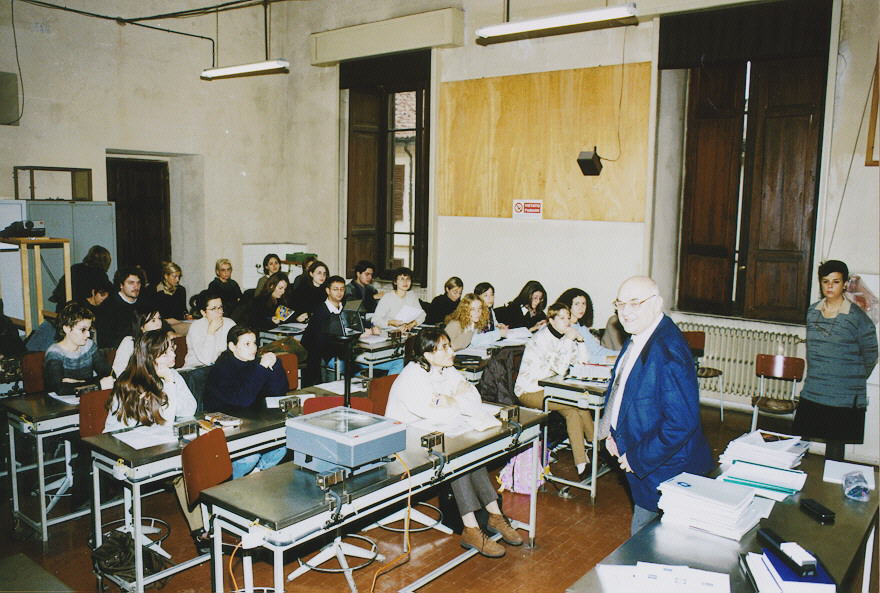
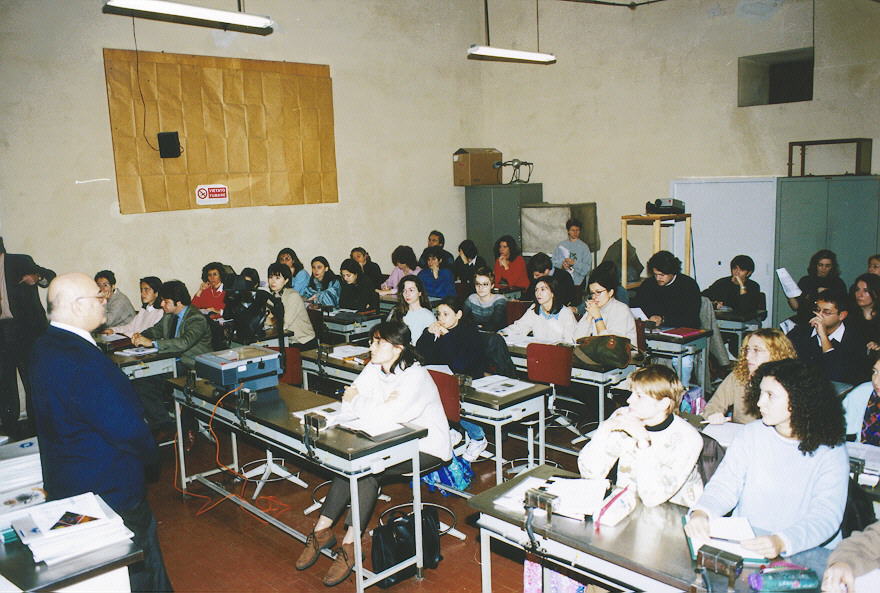
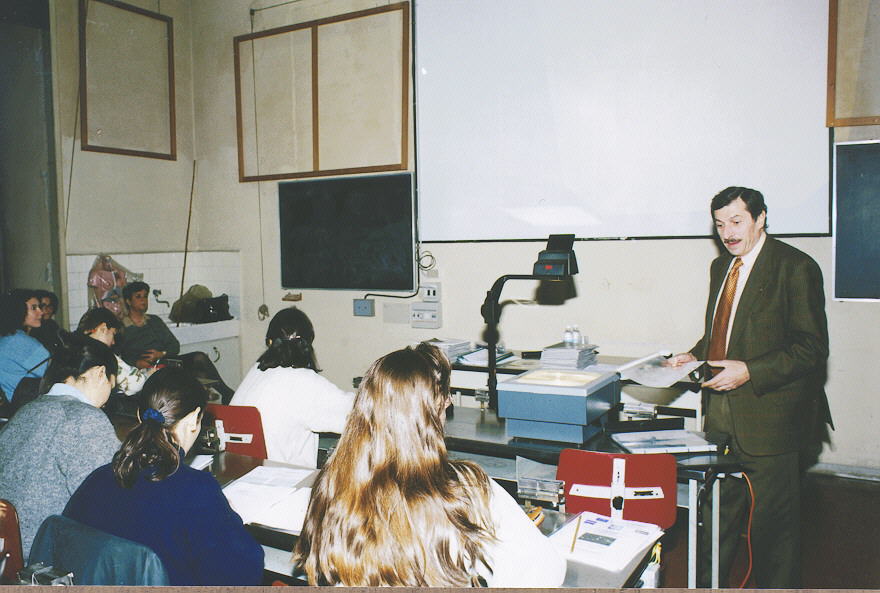
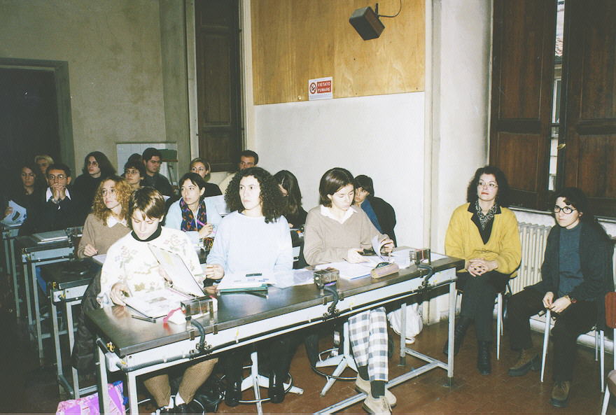
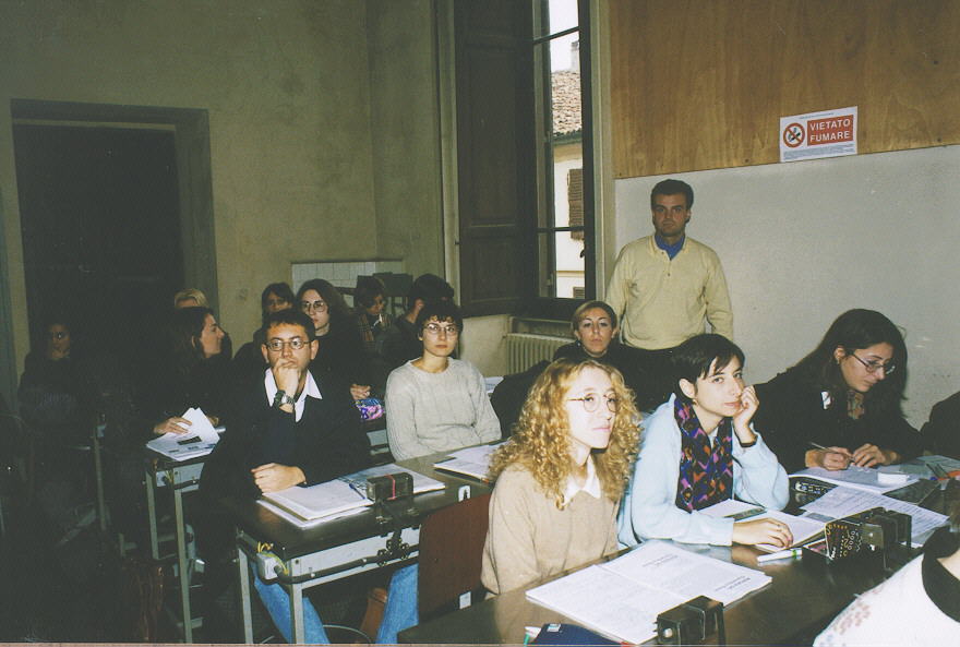
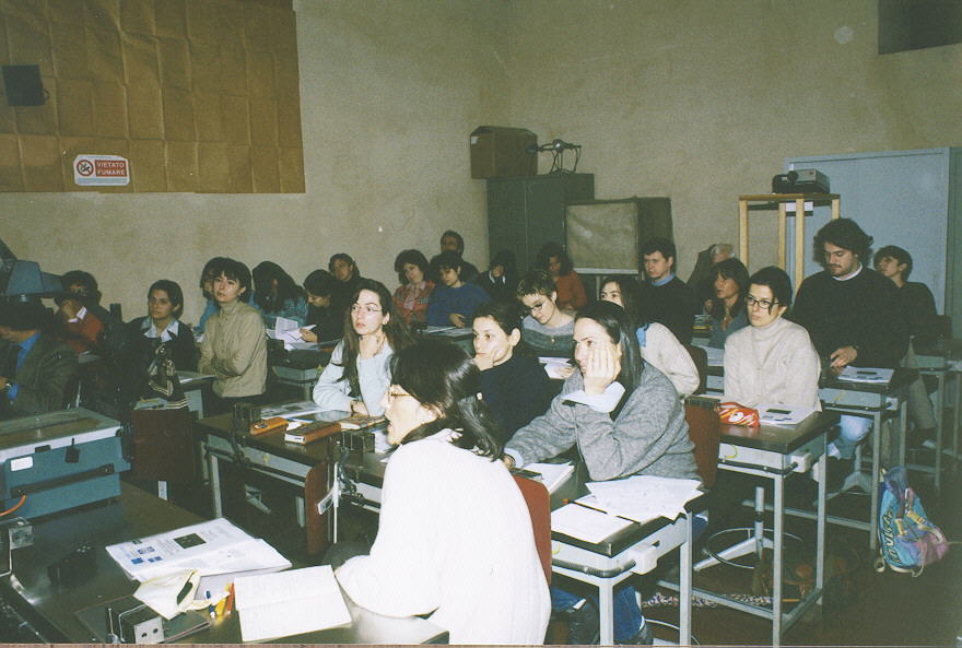
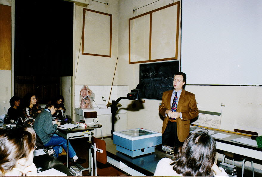
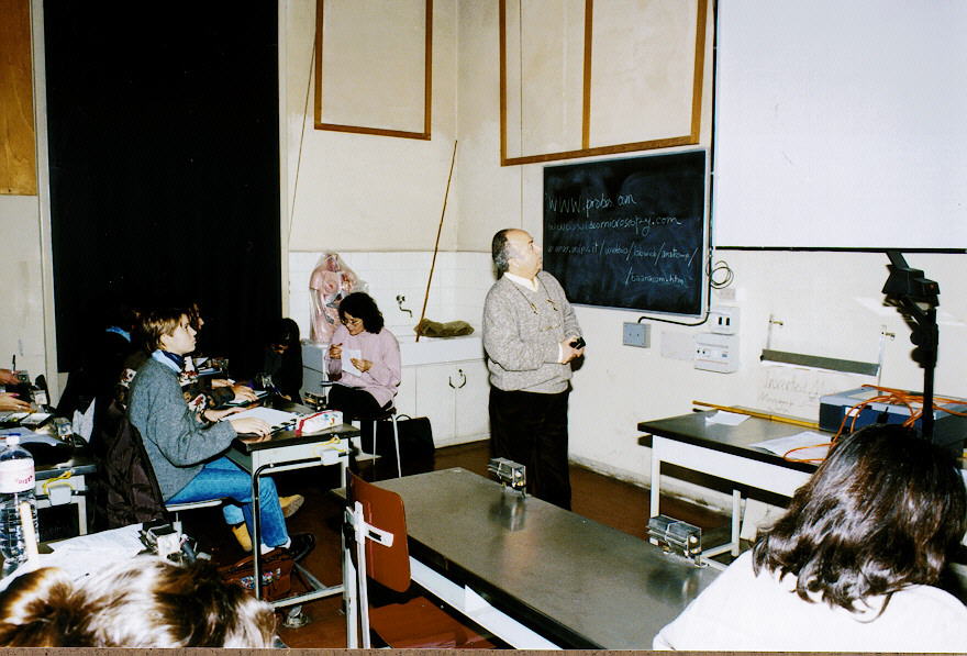
F I N A L R E P O R T
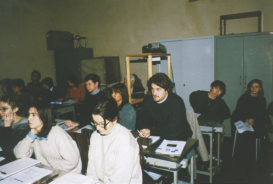
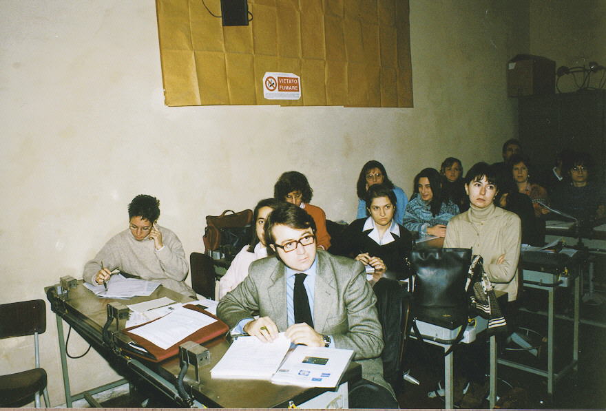
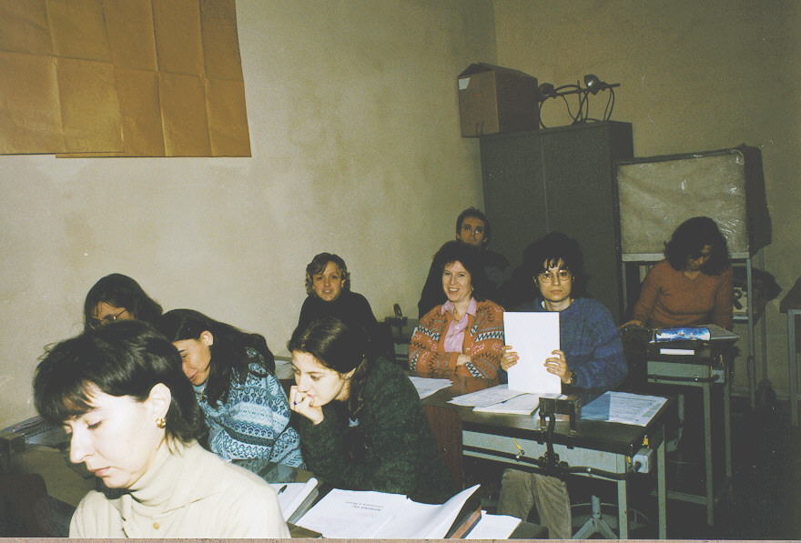
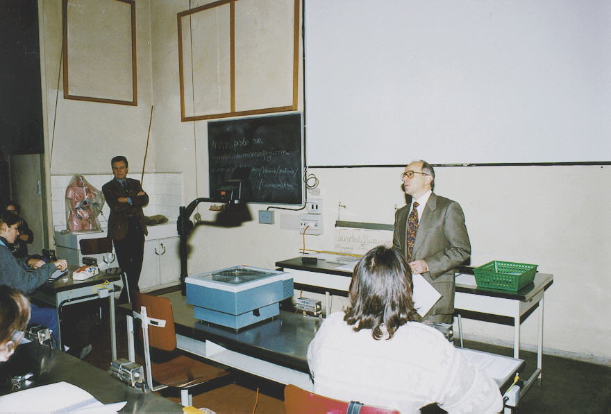
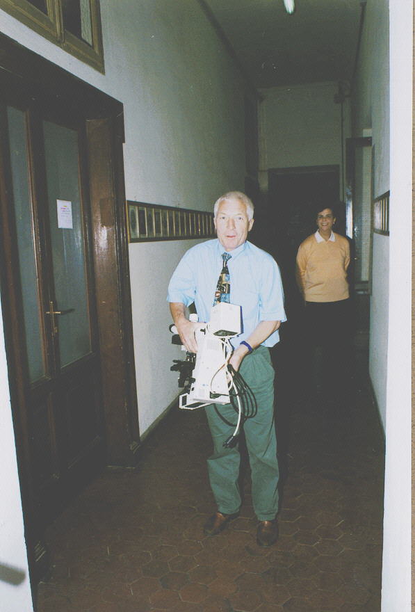
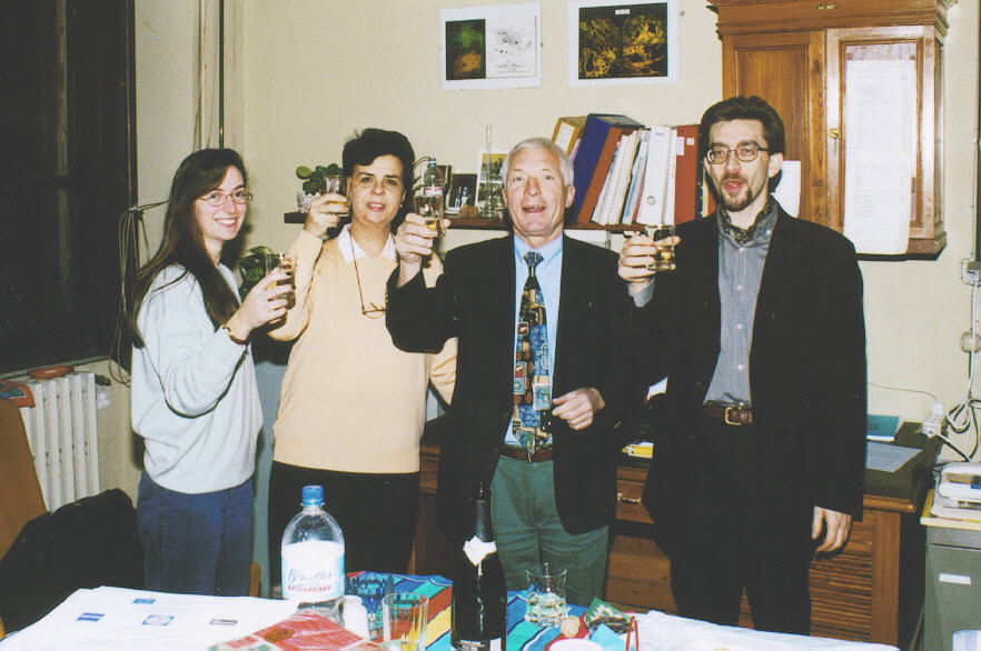
The continuing education course "Basics in fluorescence
microscopy and fluorochromes" held in the 26 and 27th november 1998
in the Department of Animal Biology of the University of Pavia, was
attended by 52 researchers in the biomedical field, coming from the Universities
of Pavia, Milan and Turin. The theoretical notions imparted in the morning
tutorials by Drs. Bottiroli and Mazzini (CNR Center for Histochemistry,
Pavia), Prof. Barni (University of Pavia) and Dr. Gundlach (Carl
Zeiss, Oberkochen, Germany) were followed in the afternoon by practical
workshops during which the partecipants were allowed to observe under the
supervision of Dr. Gundlach fluorochromized samples of fixed or supravitally
stained cells on microscopes provided by Carl Zeiss SpA, Italy. Multicolor
labeling experiments were made on living cells and tissue sections using
fluorescence probes provided by Molecular Probes. The partecipants could
select single or multibandpass filters in order to obtain the best observation
conditions for each sample.
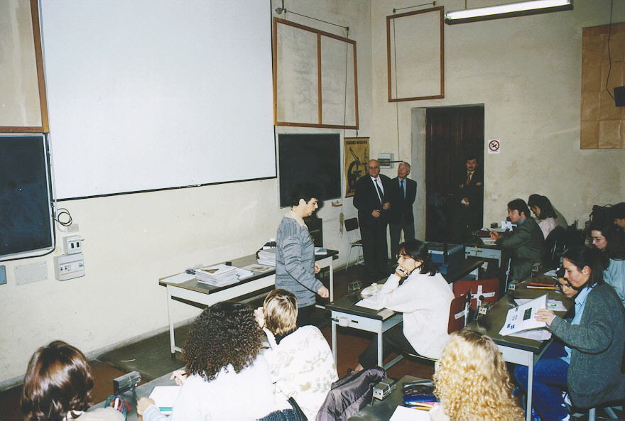
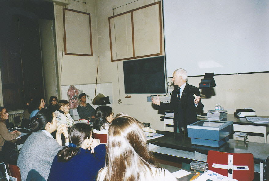
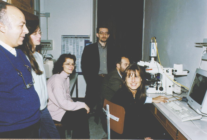
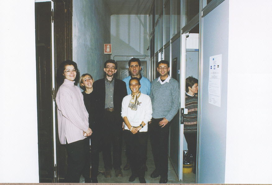
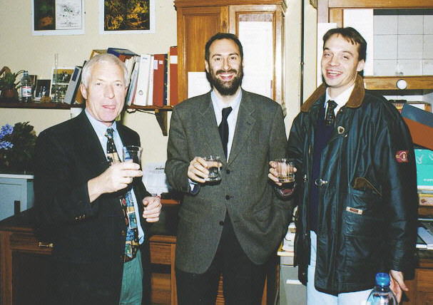
These two days were preceded by a short
training course of three days on supravital fluorescence techniques, held
by experts from the Universities of Pavia (Prof. Barni) and Sassari (Drs.
Sciola and Spano), and addressed to 5 future tutors of the Leonardo Courses
(Drs. Griffini, Bertone, Bono, Fracchiolla and Garda).
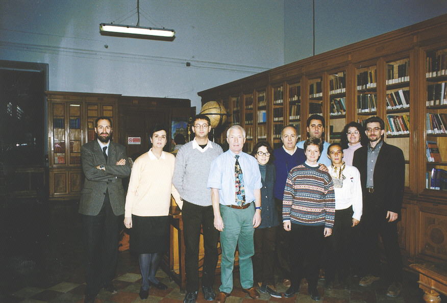
The Course organizers (Prof.s Freitas,
Pellicciari and Gundlach) acknowledge that the enormous success of both
courses was made possible due to the "back stage" efforts and hard work
of the experts Sciola and Spano, the tutors, the technicians from the Laboratory
of Cell Biology (Dr. Bottone, Mrs. Veneroni), from Carl Zeiss SpA, Italy
(drs. Borsatti and Beller) and the secretaries of the Laboratories of Comparative
Anatomy and of Cell Biology (Mr. Sanga and Mrs. Bosini).
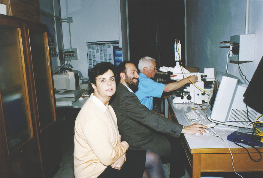
The didactic material was kindly provided
by the Gruppo Italiano di Citometria. The digital acquisition and elaboration
of the images was made by Drs. Borsatti and Bertone. The Internet advertisements,
the electronic elaboration of the booklet with didactic material and the
photographic documentation were made by Dr. Bertone.
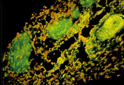
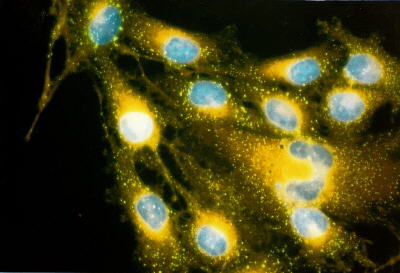
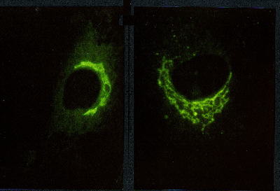
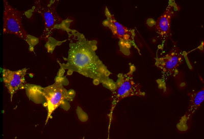
Selected pictures of the
biological samples
>> Click on
the images to see at full screen <<
Real color images: scans
from color slides
 |
 |
 |
 |
 |
|
|
|
|
|
|
|
|
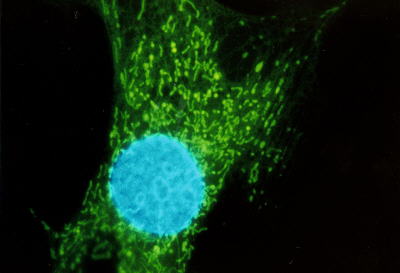
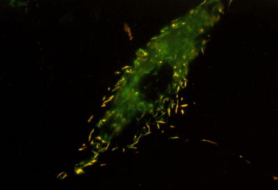
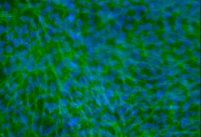
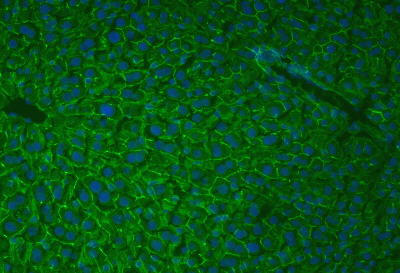
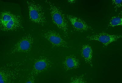
Pseudo color images: B
& W CCD SensiCam PCO camera, Germany; Axiovision software and
Axioplan II microscope, Carl Zeiss, Germany
 |
 |
 |
 |
|
|
|
|
|
|
|
|
||||||||||||||||||||||
|
This site is created and maintained by Vittorio
Bertone
Last version 3 December 1998.